MESA
Adnexal Mass Segmentation in US images
- DINOv3
- Segmentation
- US
- venue
- CVPR 2026 ⋅ SVC Workshop
- year
- 2026
Abstract
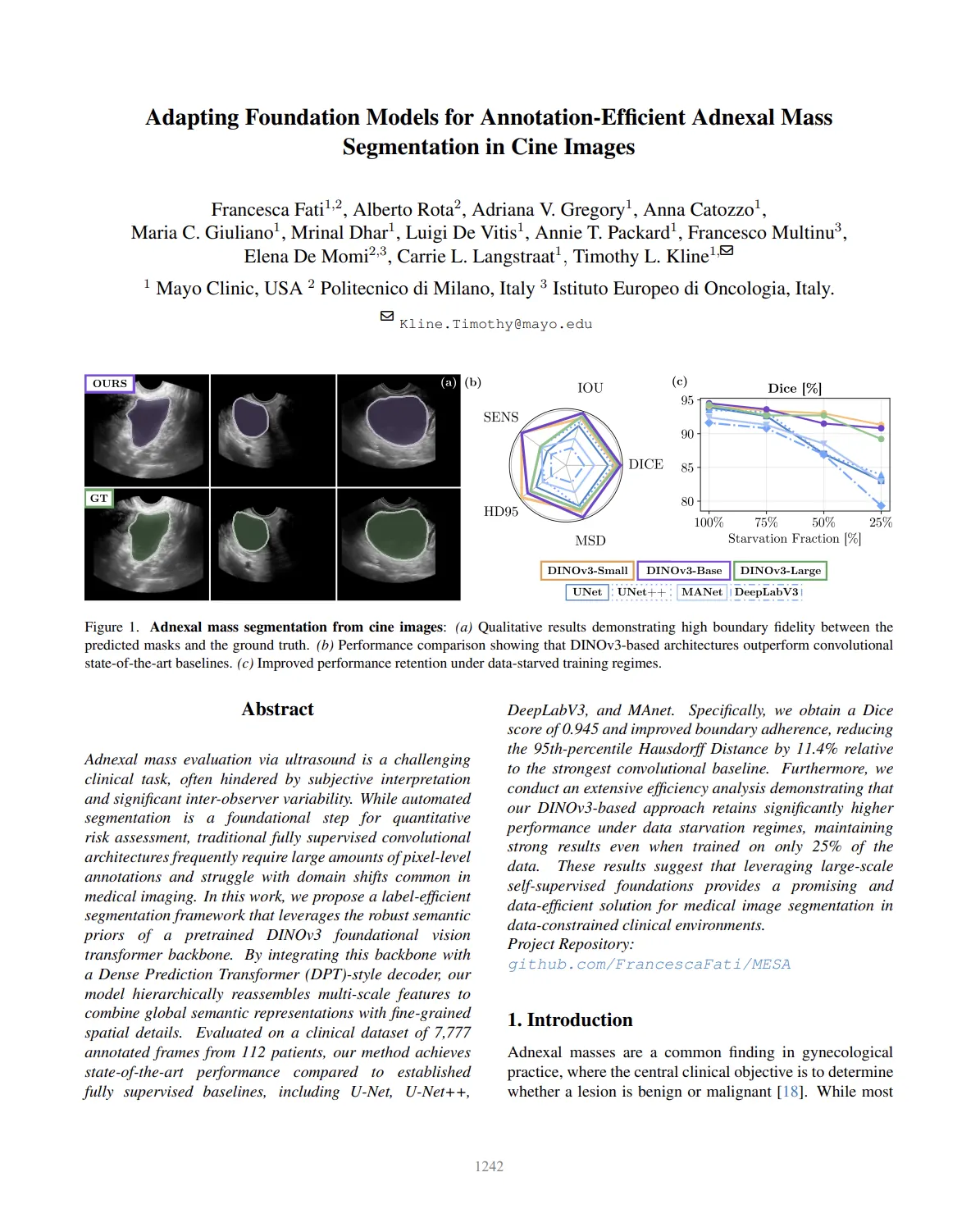
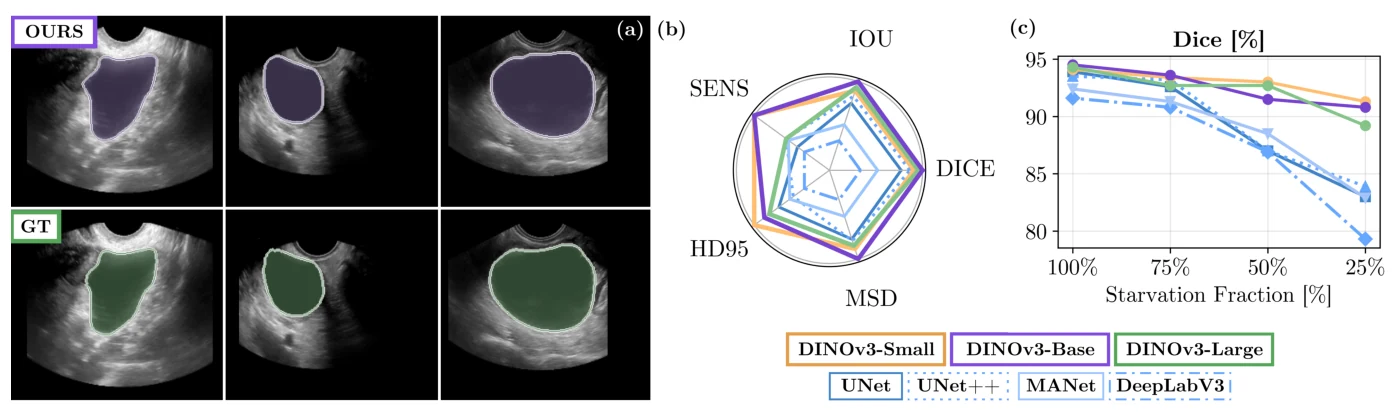
Adnexal mass evaluation via ultrasound is hindered by subjective interpretation and inter-observer variability. While automated segmentation supports quantitative risk assessment, fully supervised convolutional architectures require large pixel-level annotation budgets and struggle under domain shift. We propose a label-efficient framework leveraging a pretrained DINOv3 foundation backbone integrated with a Dense Prediction Transformer (DPT)-style decoder that hierarchically reassembles multi-scale features. Evaluated on 7,777 annotated frames from 112 patients, the method achieves a Dice score of 0.945 and reduces the 95th-percentile Hausdorff distance by 11.4% relative to the strongest convolutional baseline, while retaining strong performance when trained on only 25% of the data.
Introduction
Adnexal masses are common in gynecological practice, where the central objective is to distinguish benign from malignant lesions. Diagnostic performance depends on acquisition quality and operator expertise, leading to substantial variability. Ultrasound images are further challenged by speckle noise, acoustic shadowing, and low contrast. Accurate segmentation is a prerequisite for quantitative assessment pipelines including the IOTA ADNEX model and O-RADS risk stratification. Foundation models pretrained on large-scale natural-image corpora offer semantic priors that may transfer to medical ultrasound with minimal task-specific pretraining.
Methodology
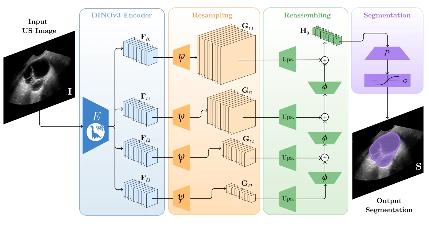
Grayscale cine frames are mapped to pseudo-RGB via channel replication and passed through a frozen DINOv3 encoder; a DPT-style decoder hierarchically reassembles features from layers {F₃, F₆, F₉, F₁₂} into a binary segmentation mask. Only the decoder is trained under a combined BCE and Dice objective (λ_BCE = 0.3, λ_Dice = 0.7) with AdamW, cosine learning-rate schedule, and 224×224 inputs. Baselines include U-Net, U-Net++, DeepLabV3, and MAnet trained under identical protocols. Data-starvation experiments train on 25%, 50%, 75%, and 100% of the labeled set to assess annotation efficiency.
Results
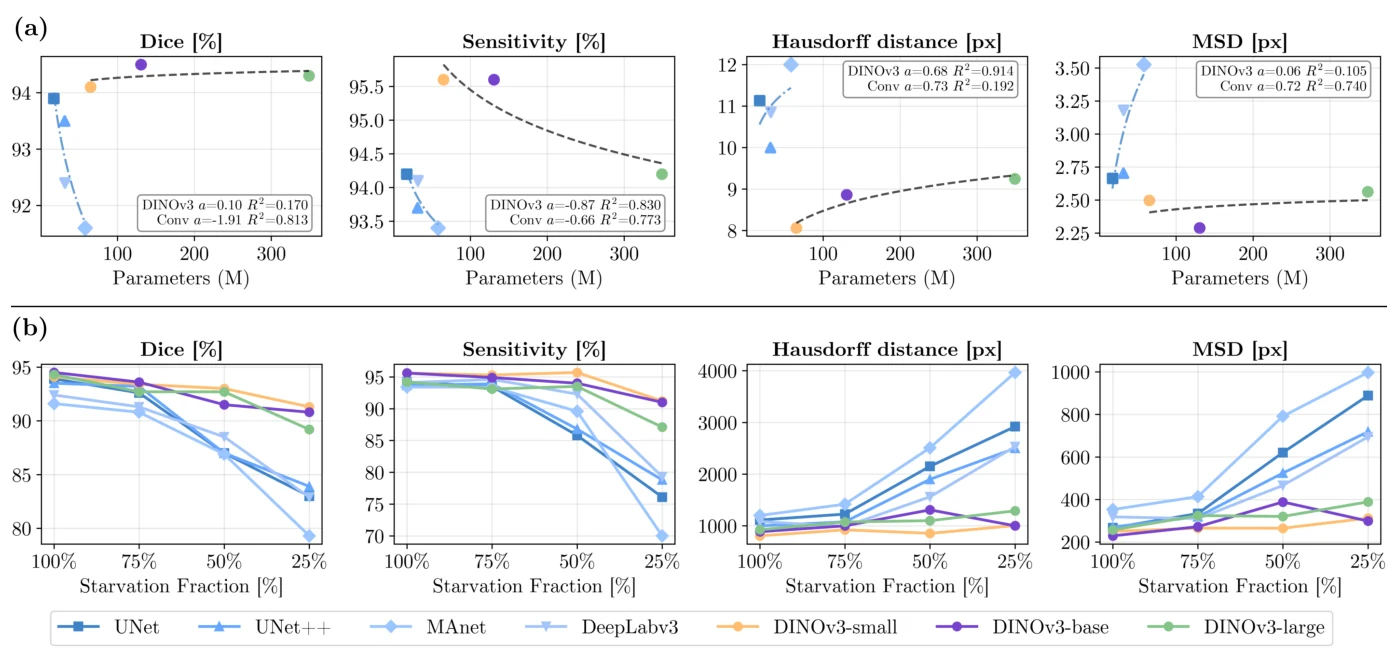
MESA reaches Dice 0.945 and the best boundary-sensitive metrics across resolutions 224×224 and 512×512, outperforming all convolutional baselines on IoU, sensitivity, and Hausdorff distance. Under data starvation, CNN architectures degrade sharply at 25% of labels while the DINOv3-based model retains substantially higher Dice and lower HD95. Ablations across DINOv3 scales (Small, Base, Large, Giant) reveal early performance saturation and overfitting at extreme scales, with moderately sized backbones offering the best accuracy–efficiency trade-off for boundary-sensitive ovarian ultrasound segmentation.
Conclusion
Integrating a self-supervised DINOv3 backbone with a DPT decoder delivers state-of-the-art adnexal mass segmentation with markedly improved contour fidelity and resilience to limited annotations. The findings support foundation-model priors as a practical path toward annotation-efficient deployment in clinical ultrasound workflows, where expert labels are costly and inter-operator variability is high.
Collaborators
- Francesca Fati
- Adriana V. Gregory
- Anna Catozzo
- Maria C. Giuliano
- Mrinal Dhar
- Luigi De Vitis
- Annie T. Packard
- Francesco Multinu
- Elena De Momi
- Carrie L. Langstraat
- Timothy L. Kline