muVES
3D analysis of microvasculature on-chip
- MATLAB
- Microfluidics
- 3D Imaging
- venue
- BTM
- year
- 2023
Abstract
Three-dimensional imaging (e.g., confocal microscopy) is commonly used to visualize microvasculature-on-a-chip models, yet 3D analysis is not standard for extracting quantitative metrics. We developed μVES to compute morphological parameters (radius, length, tortuosity, eccentricity) and intravascular flow velocity from segmented 3D volumes. Applied to fibrin-gel microvascular networks, the tool reproduces literature-consistent capillary morphology and, combined with bead-tracking velocimetry, estimates flow velocities and wall shear stresses that agree with experiments under imposed pressure gradients.
Introduction
Microfluidic microvasculature-on-a-chip models study angiogenesis, tumor extravasation, immune recruitment, and drug delivery with increased physiological relevance from human-derived cells and 3D architecture. Perfusion under controlled pressure enables functional readouts, but most morphological analyses still rely on 2D projections of confocal stacks, discarding information along the vessel axis and mis-estimating radius and surface-to-volume ratios when cross-sections are non-circular.
Methodology
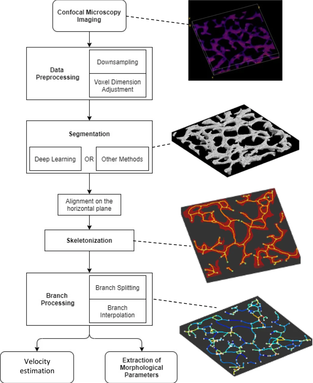
μVES comprises eight steps: acquisition, preprocessing (cubic voxel interpolation, optional downsampling), segmentation (active contours or a deep-learning alternative), network alignment, skeletonization, branch splitting, per-branch morphometry, and Poiseuille-based flow solving over the vascular graph under an assumed pressure drop. Wall shear stress is derived from velocity and local radius. A 2D projection mode (μVES2D) enables validation against REAVER and manual tracing before full 3D analysis.
Results
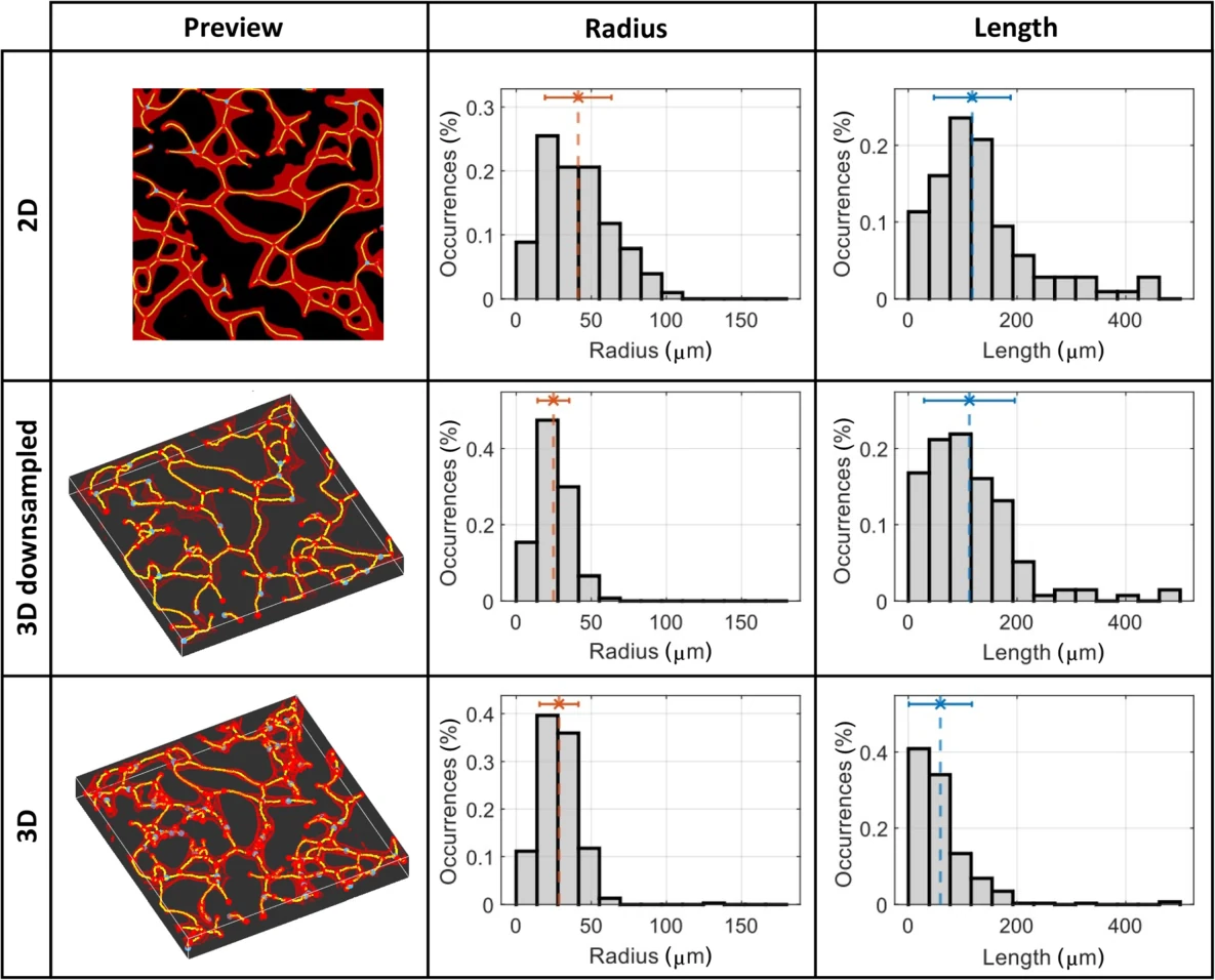
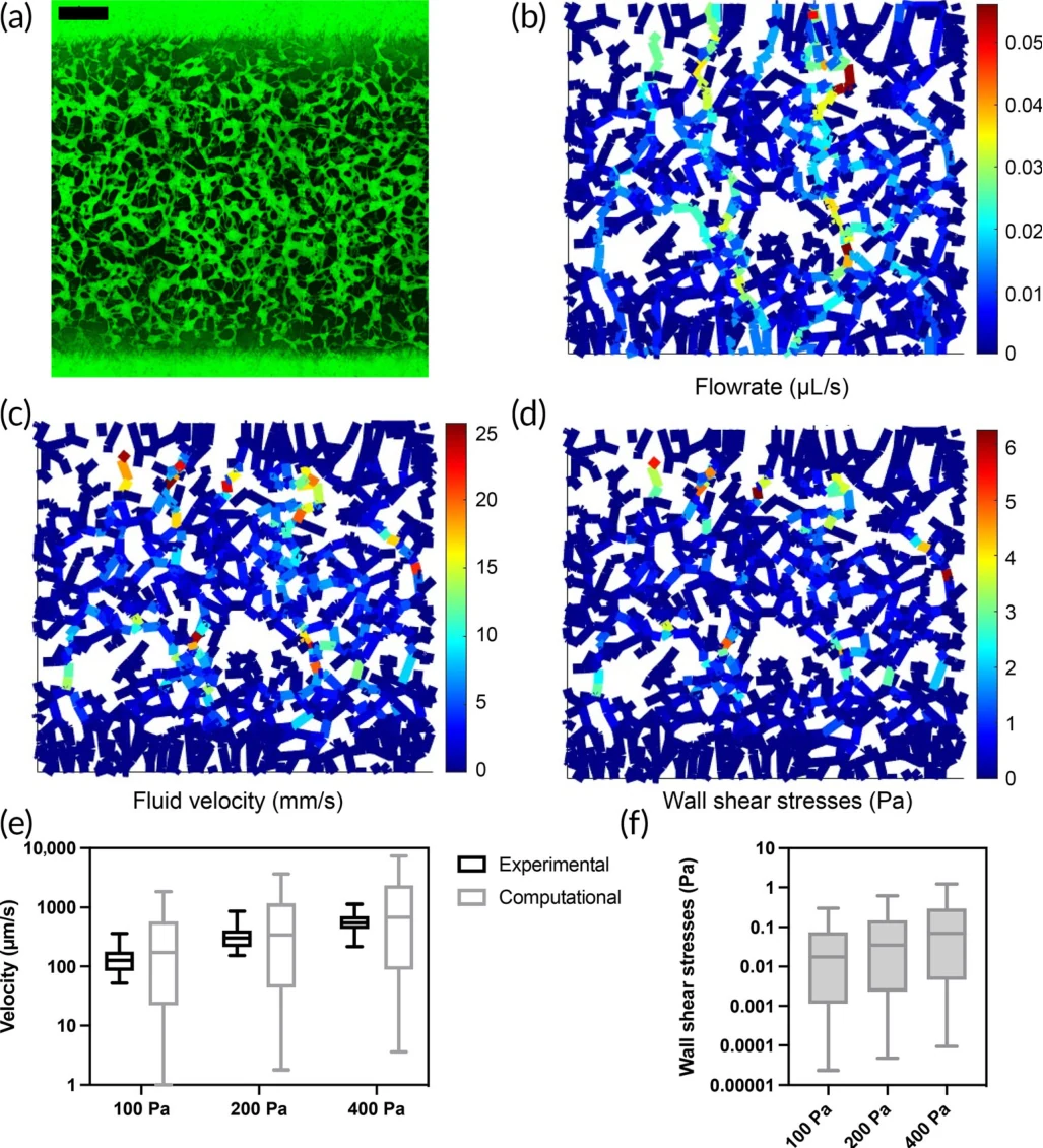
Validated against REAVER on 2D projections, μVES matches established length estimates before exploiting the full volume, where mean vessel eccentricity is 0.833 ± 0.125, revealing markedly non-circular cross-sections invisible to 2D analysis (2D mean radius 41.3 μm vs. 3D 28.5 μm on a representative network). Across 30 microvascular networks, mean capillary diameter is 31.4 ± 16.2 μm and tortuosity 1.18, consistent with in vitro literature. Flow velocities under 100/200/400 Pa pressure gradients agree with bead-tracking measurements; wall shear stress remains lower than in vivo references but approaches physiological values at higher imposed gradients. Deep-learning segmentation cuts computation time substantially with comparable morphological output.
Conclusion
μVES demonstrates that 3D analysis is essential for accurate microvascular morphometry and functional assessment in vitro. The open-source MATLAB implementation enables reproducible characterization of vasculature-on-a-chip models and highlights where current in vitro networks match or diverge from in vivo microvasculature in morphology and hemodynamics.
Collaborators
- Luca Possenti
- Giovanni S. Offeddu
- Martina Senesi
- Adelaide Stucchi
- Irene Venturelli
- Tiziana Rancati
- Paolo Zunino
- Roger D. Kamm
- Maria Laura Costantino