UnderXAI-OVIT
Decision support in ovarian cancer treatment
- PyTorch
- Explainable AI
- Medical Imaging
- venue
- Nature WH ⋅ Preprint
- year
- 2025
Abstract
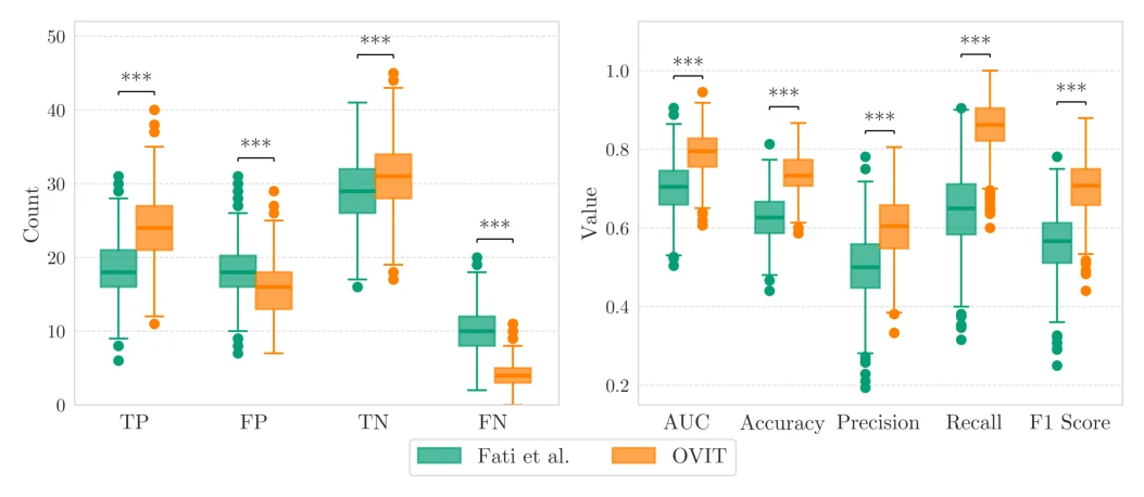
Ovarian cancer is the deadliest gynecologic malignancy, with complete resection after primary debulking surgery associated with the most favorable prognosis, yet assessing resectability at diagnosis remains challenging. We propose OVIT, a fully automated decision support system predicting residual tumor after primary debulking from preoperative CT and clinical variables, without manual segmentation or expert radiological scoring. On an independent test cohort of 75 patients from a 465-patient retrospective HGSOC cohort, the best model achieves AUC 0.80 (95% CI: 0.68–0.89) and recall 0.86 (95% CI: 0.71–0.97), correctly identifying 24 of 28 non-resectable cases including 13 of 14 intraoperatively aborted surgeries.
Introduction
High-grade serous ovarian carcinoma is often diagnosed at advanced FIGO stage III–IV with widespread abdominal dissemination. The choice between primary debulking surgery (PDS) and neoadjuvant chemotherapy followed by interval debulking is made in multidisciplinary tumor boards using criteria on anatomical dissemination and surgical feasibility, yet unforeseen unresectability discovered intraoperatively remains a critical failure mode. Automated prediction from routinely available preoperative CT and biomarkers could flag patients unlikely to benefit from PDS before surgery.
Methodology
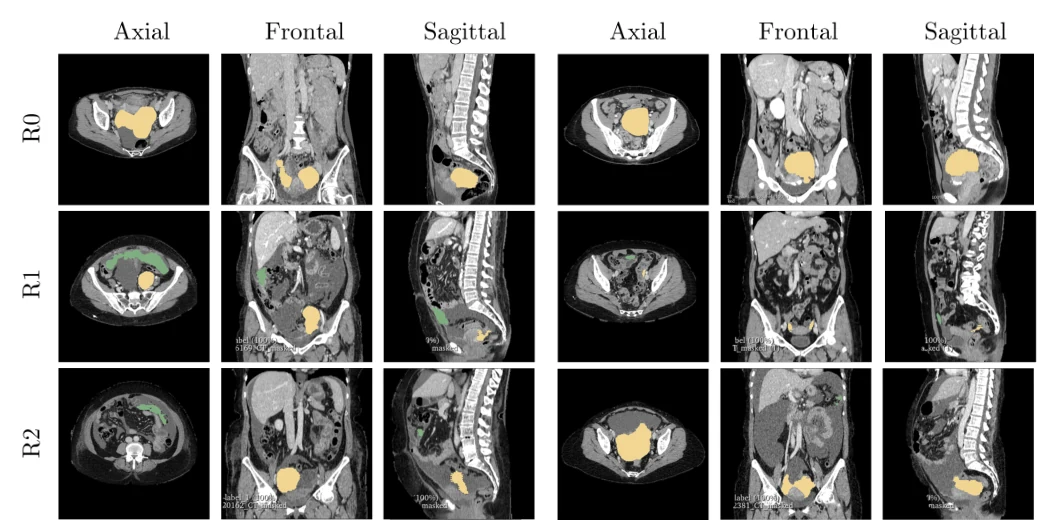
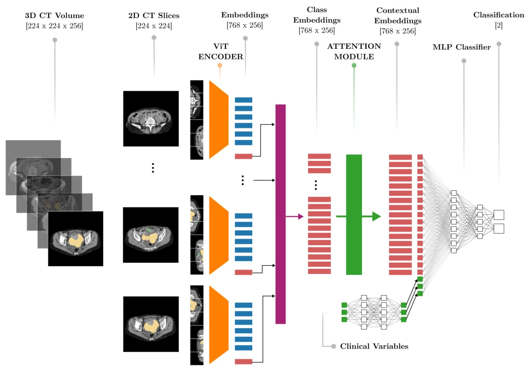
Preoperative contrast-enhanced CT volumes are automatically segmented into pelvic/ovarian and omental lesion masks by an open-source model, converted to 2D slices, and encoded slice-by-slice with a frozen pretrained Vision Transformer. Per-slice classification tokens are aggregated with an attention module and fused with clinical variables (age, CA125, HE4) through a lightweight MLP classification head; only the fusion and classification layers are trained. Four ViT backbones (DINOv2-small/base, Google-16/32) are compared with and without clinical fusion on a 390/75 train/test split from the European Institute of Oncology cohort.
Results
The Google-32 ViT encoder with clinical fusion achieves the best test performance: AUC 0.80, recall 0.86, correctly flagging 86% of non-resectable patients including 93% of surgeries aborted intraoperatively for unforeseen unresectable disease. Ablations show clinical-variable benefit is architecture-dependent: Google-32 gains most from multimodal fusion on recall-oriented metrics. Misclassifications link false negatives to older, lower-biomarker patients and false positives to elevated HE4 and higher surgical complexity scores.
Conclusion
OVIT demonstrates that frozen ViT encoders with attention pooling and clinical fusion can predict ovarian cancer resectability without manual radiological scoring. High recall on non-resectable cases supports its role as a decision-support tool to identify patients who may not benefit from primary surgery, pending prospective validation and integration into multidisciplinary workflows as part of the Under-XAI project.
Collaborators
- Francesca Fati
- Marina Rosanu
- Luigi De Vitis
- Alice Traversa
- Lucia Ribero
- Gabriella Schivardi
- Giuseppe Petralia
- Giovanni Damiano Aletti
- Nicoletta Colombo
- Michele Peiretti
- Stefano Angioni
- Jvan Casarin
- Roberto Veraldi
- Paolo Zaffino
- Maria Francesca Spadea
- Francesco Multinu
- Elena De Momi